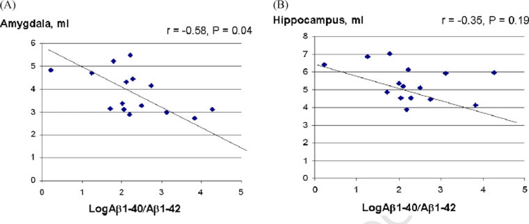
Figure 2.
Correlations between plasma Aβ1-40/1-42 and the volume of amygdala or hippocampus in amnestic MCI: The log10 of plasma Aβ1-40/1-42 ratio (x-axis) and its correlation with either amygdala volume (A) or hippocampus volume (B) (y-axis) are shown for each individual subject with amnestic MCI. Correlation coefficient factor, r, and statistical significance, p-value, are indicated.

