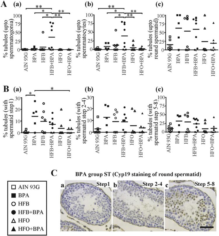
Fig. 3.
(A) The number of STs with spermatogenesis upto (a) spermatogonia (b) spermatocyte (c) round spermatid, as the last differentiated germ cell present, were tallied for male offspring exposed to the maternal diets indicated (T + E2 rodent model). (B) The number of STs with spermatogenesis impaired at the round spermatid (a) step1 (b) step 2–4 (c) step 5–8 were tallied for male offspring exposed to the maternal diets indicated. Each symbol represents a pup from an independent litter. (C) Representative CYP19 IHC staining of BPA group testis shows presence of STs with round spermatid at (a) step1 (b) step 2–4 (c) step 5–8. Bar = 40 μm. * p < 0.05, ** p < 0.01, 1-way ANOVA models between groups indicated.

