
FIGURE 2.
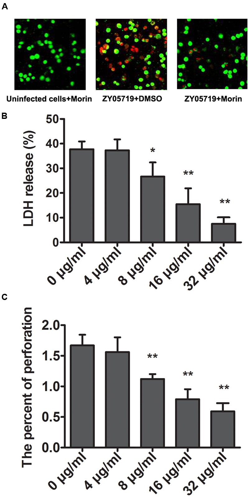
Attenuation of SS2-induced cell injury and epithelial barrier penetration by morin. (A) J774 cells were infected with SS2 in the presence or absence of morin, stained with live (green)/dead (red) agent and captured using a confocal laser scanning microscope. (B) The LDH level in the co-culture of SS2 and J774 cells treated with increasing concentrations of morin was determined using a Cytotoxicity Detection Kit (LDH) to quantify SS2-induced cell injury. Relative LDH release of each sample = (OD of tested group – OD of negative group)/(OD of positive group – OD of negative group) × 100%. (C) J774 cells were plated and cultured onto an epithelial barrier and then infected with SS2. The penetration rate of SS2 was examined by a CFU assay. ∗ indicates P < 0.05, and ∗∗ indicates P < 0.01 compared with the morin-free sample (two-tailed Student’s t-test).