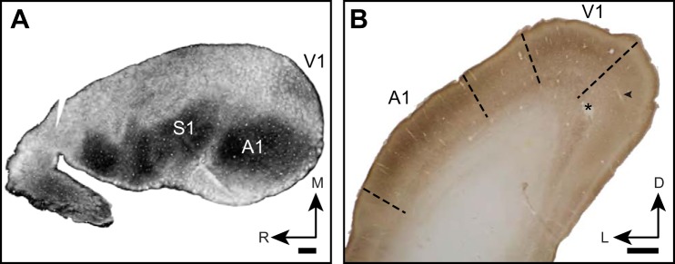
Fig. 2.
Anatomical location and identification of armadillo V1 using myelin and CO staining. A: flattened hemisphere sectioned tangential to cortical surface and stained for myelin. Note dense staining in primary somatosensory and auditory areas, while V1 is moderately stained. R, rostral; M, medial. B: photomicrograph of coronal section through caudal end of armadillo brain stained for cytochrome oxidase histochemistry. Electrode tract (arrowhead) and lesion (*) from recordings in this study are shown. L, lateral; D, dorsal. A1 and V1 are both identified by a densely stained layer IV. Relatively lightly stained region between A1 and V1 was not explored in this study but is expected to correspond to bimodal zone, in which responses to auditory and visual stimuli can be evoked. Scale bar is 1 mm.

