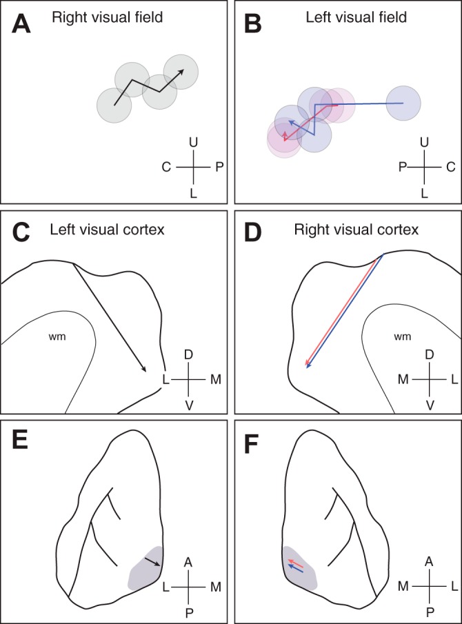
Fig. 3.

Retinotopic progression in relation to location in visual cortex. A: receptive field locations for single units recorded along electrode penetrations for case 04-16. The receptive field centers for four neurons are plotted for a single penetration, where the deepest neuron is indicated by the arrowhead. B: as in A, but for two penetrations in case 04-13. The two penetrations are indicated by red and blue symbols. Coordinates used: U, upper; L, lower; C, central; P, periphery. C and D: a sagittal drawing of the armadillo neocortex depicting the electrode trajectory for the two cases. Lines indicate the penetrations along the sagittal plane. Coordinates used: D, dorsal; V, ventral; L, lateral; M, medial. E and F: a dorsal perspective of the armadillo neocortex, where the visual cortex is indicated by the gray shading. The locations of the penetrations are indicated by the arrows. In both cases, penetrations were angled toward the medial bank, as indicated by the arrows. Coordinates used: A, anterior; P, posterior; L, lateral; M, medial.
