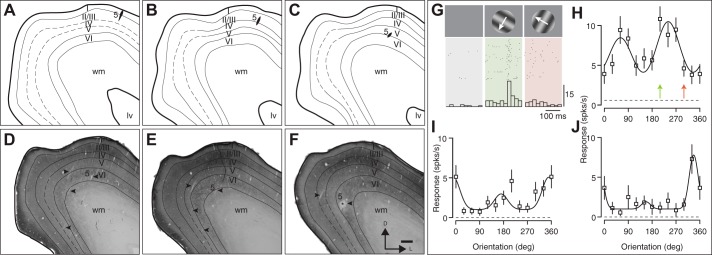
Fig. 4.
Reconstructed electrode tracks through V1 in case 14-14. A–F: drawings of sections stained for cytochrome oxidase histochemistry, spaced 100 µm apart, ordered rostrocaudally. Electrode tract for one penetration is indicated with number 5 and arrowhead (* in F indicates lesion at end of tract for penetration 5; additional arrowheads indicate other electrode tracts). In all cases, recordings were made through the cortical depths, and lesions were placed to identify the end of the penetration. Here, lv, lateral ventricle; wm, white matter. D–F include representative stained sections. Cortical layer IV was typically densely stained in V1, although in some sections, distinguishing layers III, IV, and V was difficult. G–J: data recording during penetration 5. G shows raster and histograms of the response to orientations for the orientation-selective neuron in H. Two additional neurons recorded in penetration 5 are shown in I and J. Here, spks/s, spikes per second.

