Fig. 1.

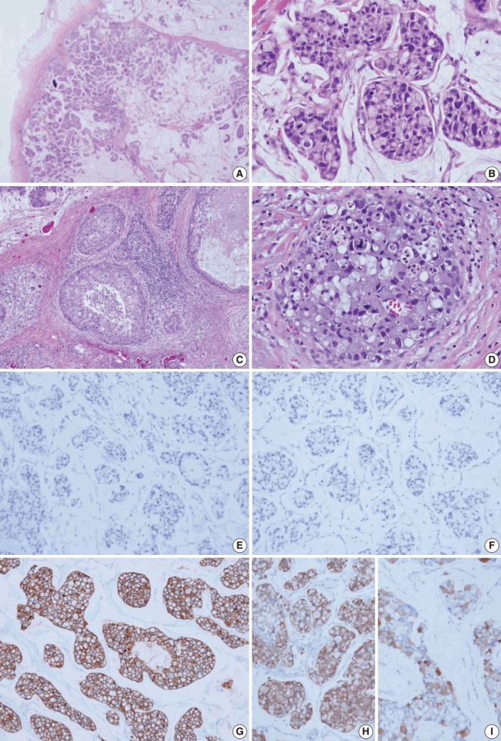
Histologic features and biomarker status in mucinous carcinoma with extensive signet ring cell differentiation. (A) In the low-power view, a tumor with an expanding margin is observed. Tumor cell clusters floating in the mucin pool are shown and the cell density is higher in the periphery than in the center. (B) In the high-power view, tumor cell cluster floating in the mucin pool shows dysplasia suitable for the nuclear grade 3. Many tumor cells are seen as signet ring cell with the nucleus pushed into the corner by abundant intracellular mucin. (C) Ductal carcinoma in situ (DCIS) is observed in the periphery of the expanding invasive nodule. (D) The DCIS component shows significantly high nuclear grade and signet ring cell differentiation. Mucinous carcinoma cells are negative for estrogen receptor (E) and progesterone receptor (F) and positive for human epidermal growth factor receptor-2 (3+) (G). Mucinous carcinoma cells are positive for mammaglobin (H) and gross cystic disease fluid protein-15 (I).
