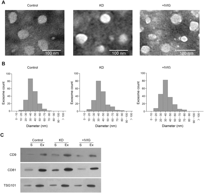
Figure 1. Characterization of serum exosomes.
(A) Typical transmission electron microscopy images of serum exosomes isolated from healthy controls (Normal), from Kawasaki disease patients (KD), and from Kawasaki disease patients after intravenous immunoglobulin treatment (+IVIG). (B) Statistics regarding vesicle diameters for the three sample types in (A), measured based on the transmission electron microscopy images. For each type of sample, 200 vesicles were measured. (C) Western blot of exosome protein markers (CD9, CD81, and TSG101) in total serum and isolated exosomes. Equal amounts of protein were loaded in each lane.

