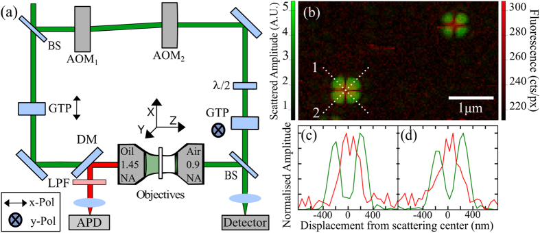
Figure 1.
(a) Schematic diagram of interferometric cross-polarization microscope (ICPM) that combines the ability to detect scattering of single nanoparticles and single molecule fluorescence, further details in main text. (b) Overlayed false colour images of scattering and fluorescent signal (green and red respectively) produced by 10 nm fluorescent nanodiamond demonstrating the excellent overlap we achieve. The applied optical power for λ = 532 nm is 37.0 μW. Images are captured at a pixel (px) resolution of 36 nm/px × 39 nm/px over an imaging area of 4.5 μm × 2.9 μm at 124 pixels × 74 pixels collected at a scan rate of 0.24 s/line. (c,d) line scans 1 and 2 for one of the fluorescent nanodiamond particles in (b).

