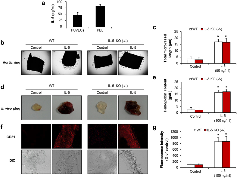
Figure 3. The neo-microvessel formation of angiogenesis in IL-5-deficient mice.
(a) ELISA immunoassay of IL-5 in cultured HUVECs. (b) Aortic ring assay in IL-5-deficient mice at 9 days. (c) Quantification of aortic ring sprouting. (d) Images of matrigel plug angiogenesis assay at 7 days. (e) Determination of hemoglobin contents in the matrigel plug. (f) CD31 staining in matrigel plugs. (g) The area of CD31-positive vessels was quantified. All data are reported as the means ± SE from three independent experiments. *P < 0.05 compared with control.

