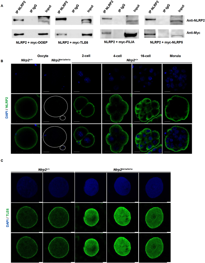
Figure 4. NLRP2 interacts with SCMC components TLE6, OOEP, FILIA and NLRP5.
(A) NLRP2 was overexpressed with myc-tagged OOEP, TLE6, NLRP5 and FILIA in HEK293T cells for 48 hours, immunoprecipitated with anti-NLRP2 or IgG as negative control and immunoblotted with anti-myc. Top panel shows specificity of anti-NLRP2 IP and bottom panel shows that NLRP2 binds to OOEP, TLE6, FILIA and NLRP5. Uncropped, full length western blots have been provided in Supplementary Figure 5. (B) Whole-mount immunofluorescence co-staining with anti-NLRP2 (green) and DAPI (blue) for nuclear staining on Nlrp2+/+ oocytes and embryos at 2-, 4-,16-cell and morula stages revealed a predominantly SCMC-like localization for NLRP2. Scale bars represent 200 μm. (C) Co-staining with anti-TLE6 (green) and DAPI (blue) of paraffin-embedded oocyte sections reveals a typical cortical stain of TLE6 in oocytes of Nlrp2+/+ dams but a more intense and diffuse stain in oocytes of Nlrp2tm1a/tm1a dams. Scale bars represent 10 μm.

