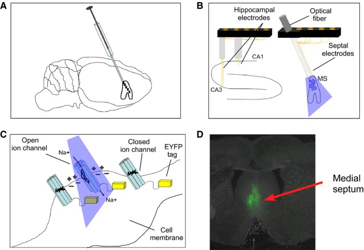
Figure 1.

Methodology of AAV injection containing channelrhodopsin (ChR2) into the medial septum, optical stimulation, hippocampal, and medial septum electrophysiology: (A) ChR2 was delivered by AAV injection into the medial septum of adult rats. Expression of ChR2 in the pace‐making cells of medial septum allows for the optical control of septal oscillations; (B) Arrangement of optical probe and EEG recording wires in the medial septum as well as EEG recording wires in both CA1 and CA3 fields of the dorsal hippocampus; (C) In response to blue light, ChR2‐expressing neurons undergo a conformational change leading to the opening of cation channel pores and the conductance of positively charged ions such as Na+. The C‐terminal end of ChR2 extends into the intracellular space and is replaced by yellow light‐sensitive proteins (EYFP indicated by yellow blocks) that were used for visualizing the morphology of ChR2‐expressing cells shown in D; (D) Histological example of forebrain tissue section under yellow light. Green fluorescence indicates cells in the medial septum expressing ChR2.
