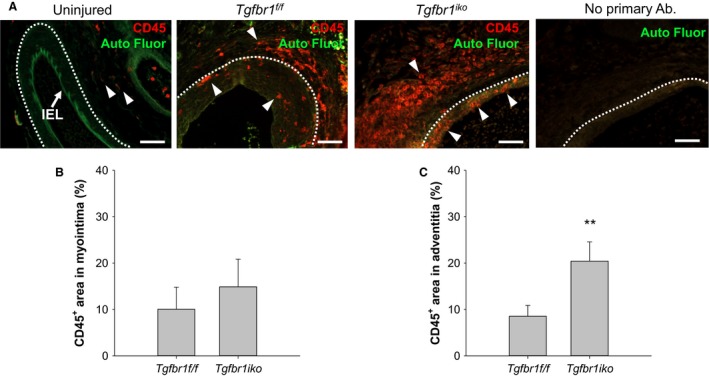
Figure 6.

Tgfbr1 iko femoral arteries (FAs) enclose more inflammatory infiltrates than do Tgfbr1 f/f FAs at the advanced stage. (A) Immunofluorescence staining of CD45 in 28‐day‐old FA lesions. An uninjured FA was included as a biological negative control (i.e., no CD45 signal in the tunica media) in the assays. A panel of “no primary antibody” negative control is shown on the far right. Red: CD45+ cells (arrowheads); green: autofluorescence; white dashed lines: EEL. Scale bar, 50 μm. Fraction of CD45+ area in the myointima (B) and adventitia (C) 28 days after injury (n = 7 per genotype). P > 0.05 (B), **P = 0.009 (C), t‐test.
