Figure 7.

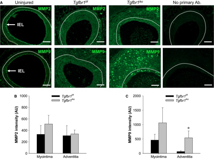
Tgfbr1 iko femoral arteries (FAs) produce higher levels of MMP9 than do Tgfbr1 f/f FAs in the adventitial layer at the advanced stage. (A) Immunofluorescence staining of MMP2 and MMP9 in FAs with indicated genotype on d28. An uninjured Tgfbr1 f/f FA was included as a reference for the baseline levels of these proteins. Positive staining of these proteins appears in green. Negative controls omitting the primary antibodies are shown on the far right. White dashed lines delineate EEL. Scale bars, 50 μm. Levels of MMP2 (B) and MMP9 (C) in myointimal and adventitial layers of the indicated FAs (n = 7 per genotype). Data were expressed as intensity of the staining (arbitrary unit, AU) normalized to the corresponding region. *P = 0.039 (C), t‐test.
