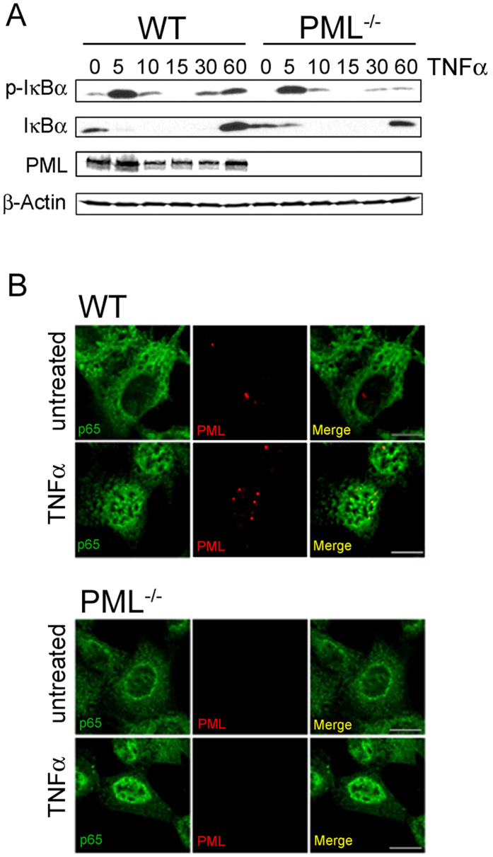
Figure 3. Normal NF-κB activation in PML−/− cells.
(A) WT and PML−/− MEFs were stimulated with TNFα (10 ng/ml) for the indicated times and analysed by immuno-blotting with the indicated antibodies. (B) Nuclear translocation of p65 in TNFα treated WT and PML−/− MEFs was assessed by confocal microscopy.

