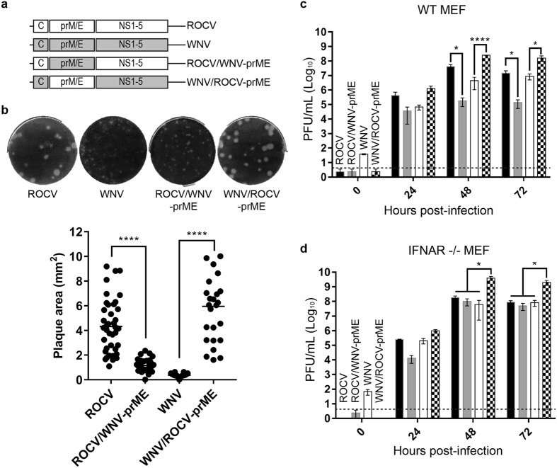
Figure 2. Construction and characterization of ROCV/WNV chimeric viruses.
(a) Schematic representation of ROCV/WNVNSW2011 chimeric viruses. (b) Plaque morphology of parental (ROCV and WNV) and chimeric (ROCV/WNV-prME and WNV/ROCV-prME) viruses in BHK-21 cells (upper) and comparison of plaque size areas (lower). Plaque areas (mm2) for 25–40 randomly selected plaques were measured using ImageJ software (National Institutes of Health, USA) and plotted as mean ± SD. (c) Growth kinetics of parental and chimeric viruses in IFN-competent mouse embryonic fibroblasts (WT MEF) and (d) IFNAR−/− MEF cells infected at MOI = 0.1. Viral titers were determined by plaque assay on BHK-21 cells at the indicated time points. The dashed lines represent the LOD of the assay. Parametric one-way ANOVA test was used to compare within groups. *P-value ≤ 0.05, ****P-value ≤ 0.0001.

