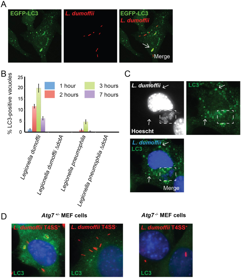
Figure 2. LdCVs become transiently decorated with LC3 in a Dot/Icm-dependent manner.
(A) Confocal z-stack of RAW cells stably expressing GFP-LC3 after infection with L. dumoffii for 3 hours. The representative LC3-positive vacuole is shown by a white arrow. (B) Quantification of LC3-association to LdCVs and LpCVs over time in THP-1 cells. Wild-type L. dumoffii capable of translocating bacterial effectors elicited LC3-decoration, whereas the translocation-deficient strains (∆dotA) did not. (C) L. dumoffiii ∆flaA in a mouse embryonic fibroblast (MEF) at 16 hours post-infection. Despite the presence of LC3-positive puncta (white arrows), significant association with the bacterial compartment (dotted white square) was not observed. (D) Representative images of LC3-association with T4SS+ and T4SS− (∆dotA) L. dumoffii in Atg7+/− MEF cells, and T4SS+ L. dumoffii in Atg7−/− MEF cells. In panels C and D, LC3 was detected by indirect immunofluorescence using anti-LC3 antibody (MBL#PM036). Red-coloured L. dumoffii constitutively express mCherry.

