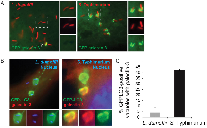
Figure 6. LC3-associated LdCVs preclude galectin-3.
(A) HeLa cells stably expressing FcγRII receptor were co-transfected with pTLR2 and either GFP-galectin-3. Images shown are at 2 hours post-infection for opsonized L. dumoffii (left) and S. Typhimurium (right), both organisms harbour plasmids for constitutive expression of mCherry (red). (B) Representative fluorescent micrographs showing co-localization of GFP-LC3 and galectin-3 on SCVs but not LdCVs. RAW264.7 cells stably expressing GFP-LC3 were infected with either S. Typhimurium (right) or L. dumoffii (left). Bacteria are shown in blue (Hoescht 3342). Galectin-3 was detected by indirect immunofluorescence using an anti-galectin-3 antibody (Santa Cruz #sc-23938). (C) Quantification of assay shown in panel B. Data are average of three independent experiments, each performed in triplicate.

