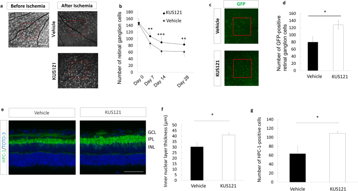
Figure 2. Effects of VCP modulators on retinal cells in an ischemic retinal injury model.
(a,b) RGC numbers in the ischemic retina using SLO imaging. (a) Representative images from animals treated with KUS121 or not 28 days after ischemia. Bar = 500 μm. (b) Longitudinal changes in RGCs counted within the red boxes in a in control (n = 11, triangles) and KUS121-treated (n = 11, circles) groups. Error bars indicate SD. **P < 0.01 and ***P < 0.005 vs. control (Student’s t test). (c and d) GFP-positive RGC numbers in flat-mounted retinas treated with KUS121 or not 14 days after ischemia. (c) Representative images. (d) GFP-positive RGC numbers counted within the red boxes in c in control (n = 8) and KUS121-treated (n = 10) groups. Error bars indicate SE. *P < 0.05 vs. control (Student’s t test). (e–g) Retinal section analysis 14 days after ischemic retinal injury. (e) Immunohistochemical images of retinal sections stained with HPC-1 and TOTO-3. Bar = 50 μm. (f) Inner nuclear layer thickness (each group, n = 8). *P < 0.05 vs. control (Student’s t test). (g) Numbers of HPC-1–positive cells in the inner nuclear layer (corresponding to amacrine cells) (each group, n = 8). *P < 0.05 vs. control (Aspin-Welch t test).

