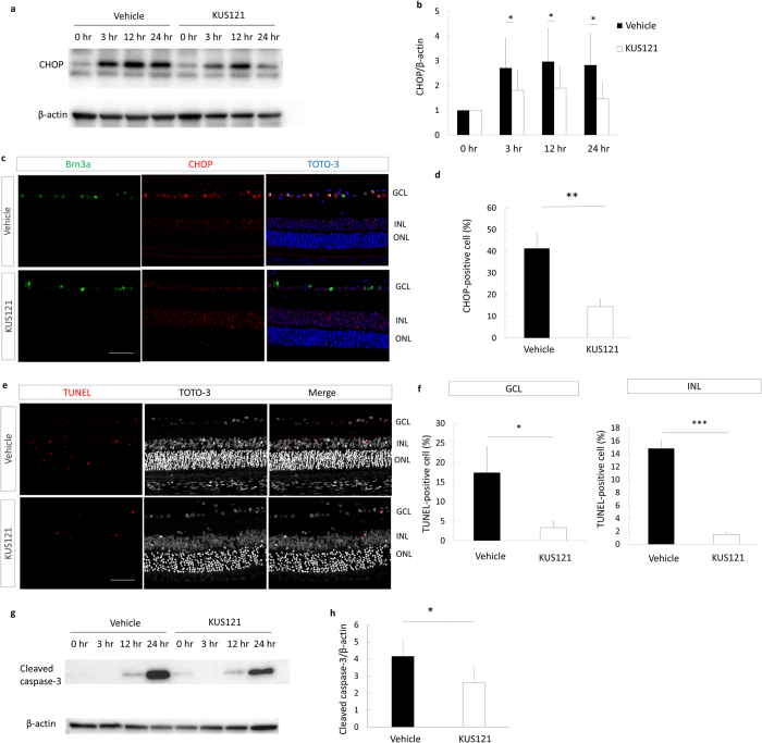
Figure 5. Mechanisms of the neuroprotective effects of VCP modulator on neural ischemia.
(a) Western blot analysis of the CHOP and beta-actin expression of rat retina before ischemic injury and 3, 12, and 24 h after ischemic injury. Ischemic injury increased the expression level of CHOP in the retina. (b) Statistical analysis of the CHOP and beta-actin expression of rat retina after ischemic retinal injury (each point of each group, n = 5), indicating that the protein level of CHOP was significantly lower in the KUS121 group than in the control group 3, 12, and 24 h after ischemic retinal injury. Error bars indicate SD. *P < 0.05 vs. control (Aspin-Welch t test). (c) Immunohistochemical images of retinal sections stained with Brn3a, CHOP, and TOTO-3 24 h after ischemic retinal injury, showing the lower percentage of CHOP-positive RGCs in the KUS121-treated rat than in the vehicle-treated rat. Bar = 50 μm. (d) Numbers of CHOP-positive cells in the ischemic retina, showing that KUS121 significantly decreased ER stress in the RGCs (each group, n = 11). **P < 0.01 vs. control (Aspin-Welch t test). (e) Representative TUNEL staining of retinal sections 24 h after ischemic retinal injury. There are fewer TUNEL-positive cells in the ganglion cell layer (GCL) and inner nuclear layers (INL) in the KUS121-treated group than in the control group. Bar = 50 μm. (f) Analysis of the number of apoptotic cells within four 780-μm squares at a distance of 500 μm from the center of the optic nerve head, showing that KUS121 significantly decreased cell apoptosis in the GCL and INL (each group, n = 8). **P < 0.05 and ***P < 0.005 vs. control (Aspin-Welch t test). (g) Western blot analysis of cleaved caspase-3 and beta-actin in the rat retina before ischemic injury and 3, 12, and 24 h after ischemic injury. Ischemic injury resulted in cleaved caspase-3 upregulation. (h) The protein levels of cleaved caspase-3 were significantly lower in the KUS121 group than in the control group at 24 h after ischemic retinal injury (each group, n = 5). Error bars indicate SD. *P < 0.05 vs. control (Aspin-Welch t test).

