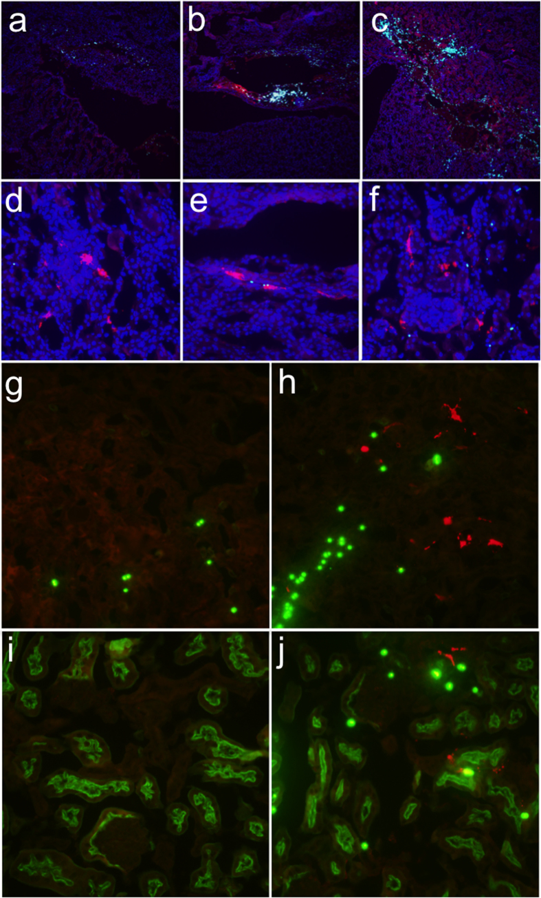
Figure 4. Renal pelvis injection results in expression in multiple kidney cell types.
Anesthetised mice were injected in the renal pelvis with a 100 μl solution containing latex fluorescent microspheres without (a) or with (b,c) 20 μg pEF-1α-TdTomato (n = 3). High concentrations of beads (small cyan circles) were found within damaged areas (a–c). In the presence of DNA, the areas of damage were colocalised with TdTomato (red) (b,c). TdTomato-positive cells in the glomerulus (d), tubules (d–f), and collecting duct (e), colocalised with beads. In (a–f) nuclei were counterstained with DAPI (blue). (g–j) Sections costained for TdTomato (red) and fluorescein lotus lectin (green) to mark the proximal tubules. Microspheres appear as bright green circles. (g) Medulla of mouse receiving beads alone. (h) Medulla of mouse receiving beads and pEF-1α-TdTomato. (i) Cortex of mouse receiving beads alone. (j) Cortex of mouse receiving beads and pEF-1α-TdTomato.

