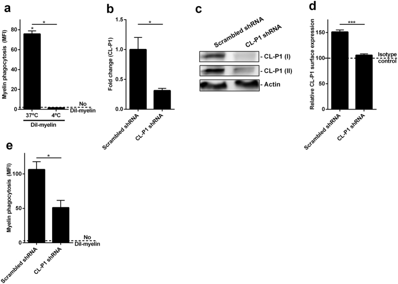
Figure 5. CL-P1 is involved in the uptake of myelin.
(a) HEK293.1 cells were exposed to DiI-labeled myelin for 1.5 h (n = 4). Myelin uptake was assessed using flow cytometry. Cells were exposed to myelin at 4°C (binding) or 37 °C (binding and uptake). Dotted line represents untreated cells. (b–d) HEK293.1 cells were exposed to scrambled shRNA or a pool of shRNA directed against CL-P1 (shRNA1-4) for 48 h. The mRNA and protein expression of CL-P1 was determined using qPCR (b, n = 4), western blot (c, CL-P1 I (R&D), CL-P1 II (Novus Biologicals), n = 3), and flow cytometry (d, n = 6). Western blots are displayed in cropped format. (e) HEK293.1 cells were exposed to scrambled shRNA or a pool of shRNA directed against CL-P1 (shRNA1-4) for 48 h. Next, DiI-labeled myelin was added for 1.5 h (n = 8). Flow cytometry was used to define myelin uptake. Dotted line represents untreated cells. Data are presented as mean ± SEM. *p < 0.05, **p < 0.01, ***p < 0.001.

