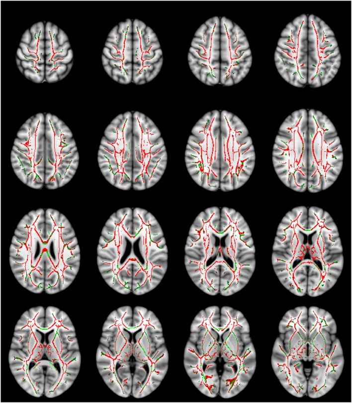
Figure 1.
Mean diffusivity (MD) values are significantly higher in the traumatic brain injury (TBI) group with normal-appearing white matter. The MD contrast is overlaid on a standard Montreal Neurological Institute 152 T1 1 mm brain and the mean fractional anisotropy skeleton (green—display threshold 0.2–0.8). The voxels where MD was found to be significantly higher in the TBI group (p-value ≤ 0.05) are shown in red. Every fourth transverse slice only is shown here to approximately cover the entire brain along the superior–inferior axis. Anatomical right side of the brain is shown on the left in this figure.

