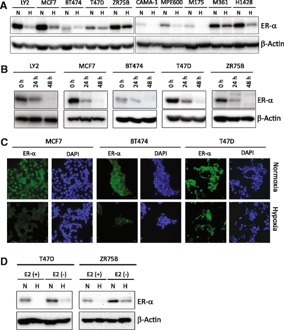
Fig. 1.

Hypoxia decreases estrogen receptor-α protein. a Representative western blot of ER-α protein from LY2, MCF7, BT474, T47D, ZR75B, CAMA-1, MPE600, MDA-MB-175, MDA-MB-361 and HCC1428 cells grown at normoxia (N) or treated with hypoxia (H)(1% O2, 24 h). b Representative western blot of ER-α protein from LY2, MCF7, BT474, T47D, and ZR75B at 24 or 48 h of hypoxia (1% O2) or normoxia (0 h). β-actin is used as a loading control. c Immune fluorescence of ER-α (green), DAPI (blue) nuclei, scale bar measures 25 μm. d Western blot of ER-α protein in ZR75B and T47D cells grown at normoxia (N) or treated with hypoxia (H) (1% O2, 24 h). E2 (+) is phenol red-free DMEM with 10% charcoal stripped FBS supplemented with 10 nM estradiol and E2 (−) is estrogen-free media (phenol red-free DMEM with 10% charcoal stripped FBS)
