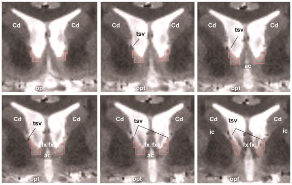
Figure 2. Delineation of the human BNST.
The BNST is identified by the red boundary (voxel size 0.8 mm × 0.8 mm × 0.9 mm) moving from the anterior (top left) to the posterior (bottom right). Cd = caudate, tsv = thalamostriate vein, ac = anterior commissure, ic = internal capsule, fx = fornix, opt = optic nerve.

