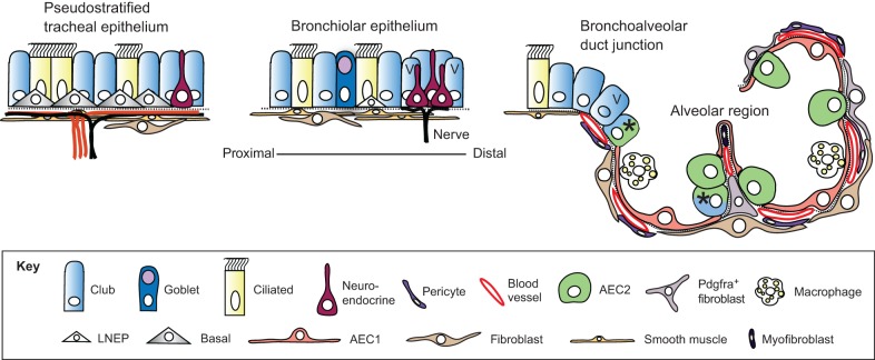
Fig. 1.
Epithelial cell types of the mouse lung. Schematic of the major cell types in different regions of the mouse lung (Hogan et al., 2014). Goblet cells are much more abundant in human versus mouse airways. Basal cells expressing detectable levels of Trp63 and Krt5 are only present in trachea and main stem bronchi. Lineage-negative epithelial progenitors (LNEPs) have been proposed for the distal airways (Vaughan et al., 2015), which in the mouse are known as bronchioles as they lack associated cartilage. There is evidence that the Club cell population is heterogeneous, with a few cells in the bronchioalveolar duct junction (BADJ) and alveoli (marked with an asterisk) co-expressing Scbg1a1 and Sftpc (Kim et al., 2005; Rawlins et al., 2009). In addition, some Club cells in the vicinity of neuroendocrine bodies and the BADJ are resistant to killing by naphthalene. These ‘variant' Club cells (marked with V) can restore the population after damage (Giangreco et al., 2002). In the alveolar region, the two major epithelial cell types are type II (AEC2) and type I (AEC1) cells. The latter are closely apposed to capillary endothelial cells. Also present are a variety of stromal cells, including Pdgfra+ fibroblasts and lipofibroblasts (the latter located close to AEC2 cells), myofibroblasts and pericytes. Image modified from Rock and Hogan (2011).

