Figure 1.
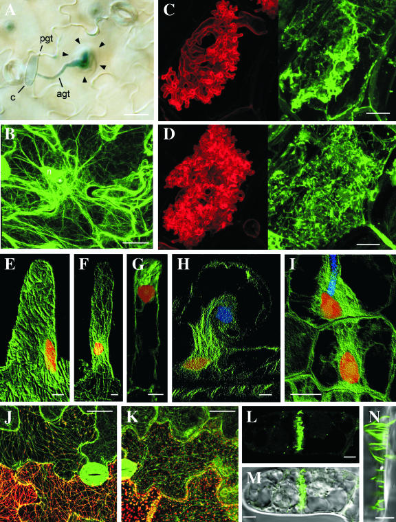
A, Accumulation of cytoplasm in an Arabidopsis epidermal cell around the attempted penetration site of the nonpathogen, B. graminis f. sp. hordei. Bar = 20 μm. B, GFP-tagged actin microfilaments focusing on the penetration site of B. graminis f. sp. hordei in an Arabidopsis epidermal cell. The fine actin microfilament network beneath the penetration site is likely to be indicative of active exocytosis. The asterisk indicates the attempted penetration site of the nonpathogen. Bar = 20 μm. pgt, Primary germ tube; agt, appressorial germ tube; c, conidum; n, plant nucleus. C and D, Arbuscules of G. versiforme developing in cortical cells of M. truncatula labeled with wheatgerm agglutinin (left) and with anti-tubulin (right). During early development (C), a diffuse fluorescence of anti-tubulin occurs around the developing arbuscular branches. Later in development (D), a dense array of short microtubules lines the perifungal membrane around the arbuscules. Bars = 10 μm. (Reproduced with permission from Blancaflor et al., 2001). E to I, Anti-tubulin labeling of microtubules in root hairs of M. truncatula before (E) and after (F–H) inoculation with rhizobia and in cortical cells (I). The helical array of cortical microtubules in the uninoculated hair (E) is replaced by a dense array connecting the nucleus with the tip of the hair (F and G) or the tip of the infection thread (H). Before the infection thread penetrates the cortical cells, a bridge of cytoplasm, the preinfection thread, forms in line with the advancing infection thread (I). Microtubules, nuclei, and infection threads are shown in green, red, and blue, respectively. E to H, Bars = 5 μm; I, bar = 15 μm. (Reproduced with permission from Timmers et al., 1999). J and K, Localization of wild-type TMV MP (J) and TMV MPR3 (K). Wild-type MP and MPR3 are visualized by tagging with DsRed (red) in transgenic plants expressing GFP-labeled microtubules (green). Bars = 25 μm. (Reproduced with permission from Gillespie et al., 2002). L to N, Fluorescent tubules in a young cross wall of a BY-2 suspension cell formed after expression of GFLV MP tagged with GFP. Bars = 5 μm. (Reproduced with permission from Laporte et al., 2003).

