
Figs. 5a - 5d.
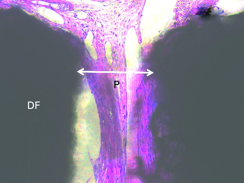
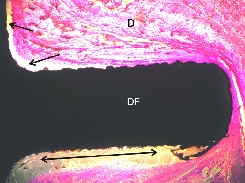
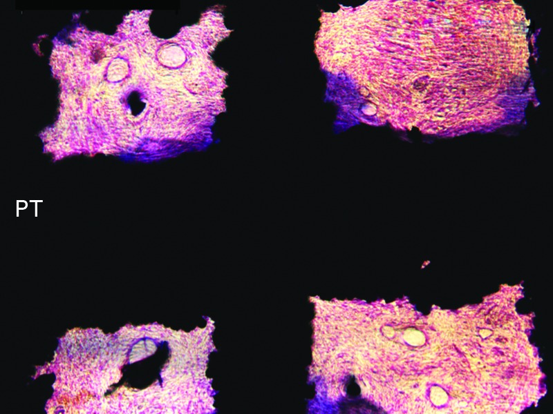

Histological sections showing (a) incomplete soft-tissue fill within a drilled flange (DF) pore (P); (b) areas of lack of the dermal tissue (D) attachment to the flange of a DF implant (arrows); (c) intimate contact between uncoated porous titanium alloy flange (PT) pore edges and soft-tissue PT. Increased collagen deposition is seen at the pore periphery and (d) the dermis attaching to hydroxyapatite with fibronectin coated flange flange.