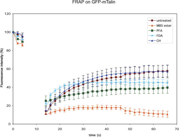
Figure 5.
FRAP reveals a dynamic flux of GFP-mTalin off and on F-actin. First three scans (single optical sections in one focal plane) were performed, whereafter a box of approximately 10 μm in length was bleached by five consecutive scans at full laser power (see Fig. 4). This interval corresponds with the data-less area in the graph. The recovery after FRAP takes place slightly slower than the cytoplasmic streaming speed (FDA control), but much faster than in cells that have been fixed with MBS-ester or paraformaldehyde (PFA). The reappearance of fluorescence continues after cycloheximide (CH) treatment, indicating that the appearing fluorescence is generated by release of existing GFP-mTalin from F-actin.

