Fig. 8.
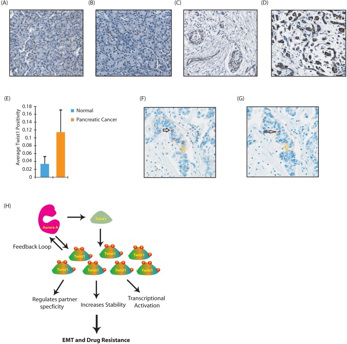
Twist1 is overexpressed in human pancreatic cancer tissues and colocalizes with AURKA. (A) Twist 1 immunohistochemistry in human normal pancreas tissues viewed at 15.0×. (B) Twist 1 immunohistochemistry in normal human pancreas tissues viewed at 15.1×. (C) Twist 1 immunohistochemistry in human pancreatic cancer tissues viewed at 15.0×. (D) Twist 1 immunohistochemistry in human pancreatic cancer tissues viewed at 15.1×. (E) Graph showing average positivity for the two groups of normal pancreas and pancreatic cancer. Pancreatic cancer had a 3-fold increase in Twist1 compared to the normal acinar pancreas. The number of tissues for the normal group and pancreatic cancer group was 5 and 12 respectively. P=0.000533 (Student's t-test for a two-tailed distribution with unequal variances). (F,G) Representative examples showing Twist1 immunohistochemistry (F) and AURKA immunohistochemistry (G) from the exact same tissue cores. These images show Twist1 and AURKA colocalization in pancreatic adenocarcinoma tissues (arrows). Imaged at 20×. (H) Proposed model showing AURKA-Twist1 axis in promoting EMT and chemoresistance in pancreatic cancer.

