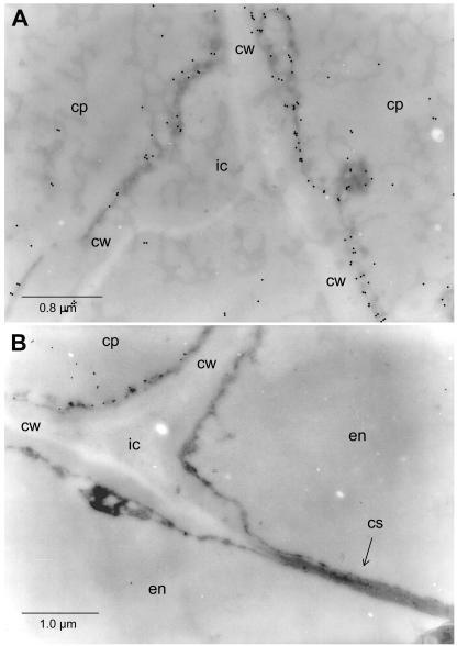
Figure 5.
Electron micrographs of in situ immunogold-labeled HSS in roots of E. cannabinum. A, Cells of the cortex parenchyma (cp) adjoining an intercellular space (ic). cw, Cell wall. B, Two cells of the endodermis (en) separated by a cell wall (cw) carrying the casparian strip (cs), an intercellular (ic), and an adjoining cell of the cortex parenchyma (cp) exclusively being labeled by the HSS-specific antibody.

