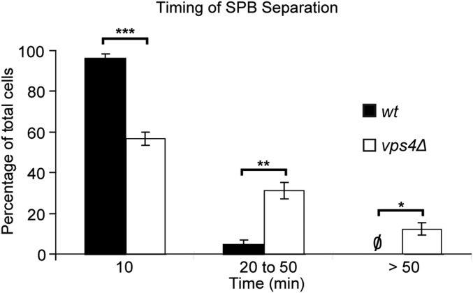
Fig. S2.
SPB separation is delayed in vps4Δ mutants. A 16-h time-lapse imaging (10-min intervals) was conducted on WT and vps4Δ cells at 32 °C, and the times required for SBP separation were scored. Data were binned and plotted by mean frequency ± SEM: WT: 10 min 96 ± 2%; 20–50 min 4 ± 2%; >50 min 0%; n = 23, 22, 22; vps4Δ: 10 min 57 ± 4%; 20–50 min 31 ± 4%; >50 min 12 ± 3%; n = 30, 30, 30. *P < 0.05; **P < 0.01; ***P < 0.001.

