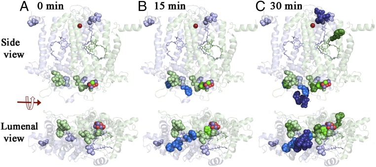
Fig. 4.
Oxidative modifications of the D1 and D2 proteins. Time course for the appearance of oxidative modifications of the D1 and D2 proteins presented as side and luminal views. The D1 protein is shown in pale green and the D2 protein is shown in pale blue. Oxidized residues are shown as spheres. Those present at 0 min are shown as pale green (D1) and pale blue (D2). Residues oxidatively labeled at 15 and 30 min are shown in progressively darker shades of green (D1) and blue (D2), respectively. The Mn4O5Ca cluster and the nonheme iron are shown as spheres. PheoD1 and QA are rendered as pale green and PheoD2 as pale blue sticks, respectively. The protein structure is from spinach PSII (PDB ID code 3JCU; ref. 6) as rendered in PYMOL (51).

