Fig. 2.
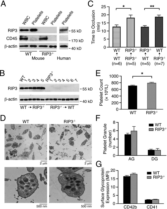
Characterization of platelets from RIP3-deficient mice. (A) Whole blood and platelets were prepared as described in SI Materials and Methods. The washed platelets and leukocytes were lysed and probed by immunoblotting with antibodies against RIP3 and CD45 (marker for leukocytes). Blotting to β-actin was used as a lane loading control. The figures are representative of three independent experiments. (B) Expression of RIP3 in platelets from RIP3−/− (WT→RIP3−/−; n = 5) and WT (RIP3−/− →WT; n = 7) mice repopulated with WT and RIP3−/− donor bone marrow- derived cells, respectively, was detected by Western blot analysis with anti-RIP3 antibody. Platelets from WT and RIP3−/− mice were used as controls, and β-actin served as a lane loading control. (C) Occlusion time of the mesenteric arteriole was measured by FeCl3 injury in WT and RIP3−/− mice repopulated with either WT or RIP3−/− donor bone marrow- derived cells. Data are mean ± SEM values. n = 5∼7. *P = 0.035; **P = 0.0047. (D) Electron microscopy analysis of WT and RIP3−/− mouse platelets. Representative images were obtained at 1,000× and 10,000×, respectively. (E) Platelet counts were detected in whole blood from WT and RIP3−/− mice (n = 26, equivalent numbers of males and females). Results are expressed as mean ± SEM. *P < 0.05. (F) Quantification of α-granules (AG) and dense granules (DG) was calculated from 20 platelets of five different fields of view for each genotype. Data are expressed as mean ± SEM. (G) Surface expression of the major glycoproteins on WT and RIP3−/− mouse platelets on membranes determined by flow cytometry with specific antibodies. Data are expressed as the mean fluorescence intensity (MFI) ± SEM of three different experiments.

