Fig. 6.
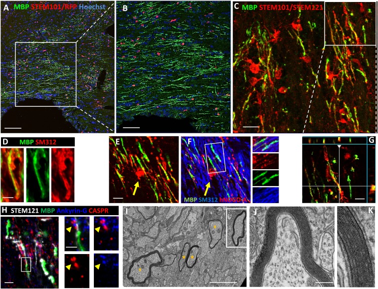
iOL give rise to functional myelin following engraftment in brains of newborn mice. (A) Transplantation of iOL into the corpus callosum of newborn Shi/Shi Rag2−/− mice resulted in extensive generation of MBP+ myelin (green) by human cells expressing RFP and staining positive for the human nuclei marker STEM101 (red) 16 wpg. (B) Higher magnification of the boxed area in A. (C) Human OL revealed by combined human nuclei STEM101 and cytoplasmic STEM121 (red) markers send multiple processes connected with MBP+ myelin. Orthogonal view of the boxed area illustrating the colocaliziation of cytoplasmic STEM121 with MBP is depicted in G. (D) Colabeling of MBP (green) and axonal neurofilament (SM312, red), highlights wrapping of host axons by donor-derived myelin. (E and F) Mature human NOGO-A+ oligodendrocyte (red, yellow arrow) connected to MBP+ myelin (green) wrapped around host axons (blue). The small panels in F illustrate the merged and single fluorochromes images represented in the boxed area. Note that unlike MBP, NOGO-A is expressed in the cell cytoplasm (cell body, processes, and paranodal loops) but not in compact myelin. (H) Human-derived (STEM121+, white)/MBP+ myelin (green) integrate into axo-glial elements expressing Ankyrin-G nodal protein (blue, yellow arrow) and CASPR paranodal proteins (red). The boxed area is enlarged to illustrate a typical node defined by a STEM121+ grafted cell and its MBP+ myelin internode, with Ankyrin-G+ aggregate flanked by two CASPR+ domains. (I–K) EM images demonstrate that human-derived myelin undergoes final maturation via compaction and formation of the major dense line. Axons surrounded by compact myelin are indicated by yellow stars. J and K are higher magnifications of boxed axon in I. n = 4 for immunostaining, n = 3 for EM. [Scale bars, 100 µm (A), 50 µm (B), 20 µm (C), 5 µm (D), 10 µm (E and F), 5 µm (H), 2 µm (I), and 200 nm (J).]

