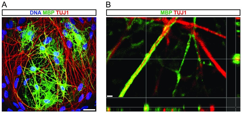
Fig. S7.
Confocal analysis of in vitro myelination assays. (A) Confocal immunofluorescence image of iOL cocultured with iPSC-derived neurons for 21 d. The image illustrates the colocalization of MBP (green) with neuronal processes visualized by TUJ1 (red). Nuclei were counterstained with Hoechst. No MBP expression was detectable in control cultures. (B) Orthogonal projection illustrates the formation of MBP+ (green) sheaths around TUJ1+ (red) neuronal processes. (Scale bars, 25 µm in A and 1 µm in B.)

