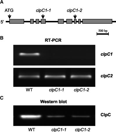
Figure 1.
Confirmation of the T-DNA insertion lines for clpC1. A, Schematic picture of the genomic clpC1 gene in Arabidopsis. Gray boxes and black lines represent exons and introns, respectively. Arrows indicate the ATG start codon and the proposed T-DNA insertion sites for the two independent mutant lines, clpC1-1 and clpC1-2. B, RT-PCR analysis of clpC1 and clpC2 gene expression of wild type (WT), and clpC1-1 and clpC1-2 mutant lines. Reactions were performed with equal total RNA, with the resulting RT-PCR products visualized by staining with GelStar. C, Immunoblot analysis of total ClpC protein in wild type, and clpC1-1 and clpC1-2 mutant lines. Total proteins were extracted from leaves of each plant and separated by denaturing PAGE on the basis of equal fresh weight. Total ClpC protein was detected by immunoblotting with an antibody that crossreacts with both ClpC1 and ClpC2.

