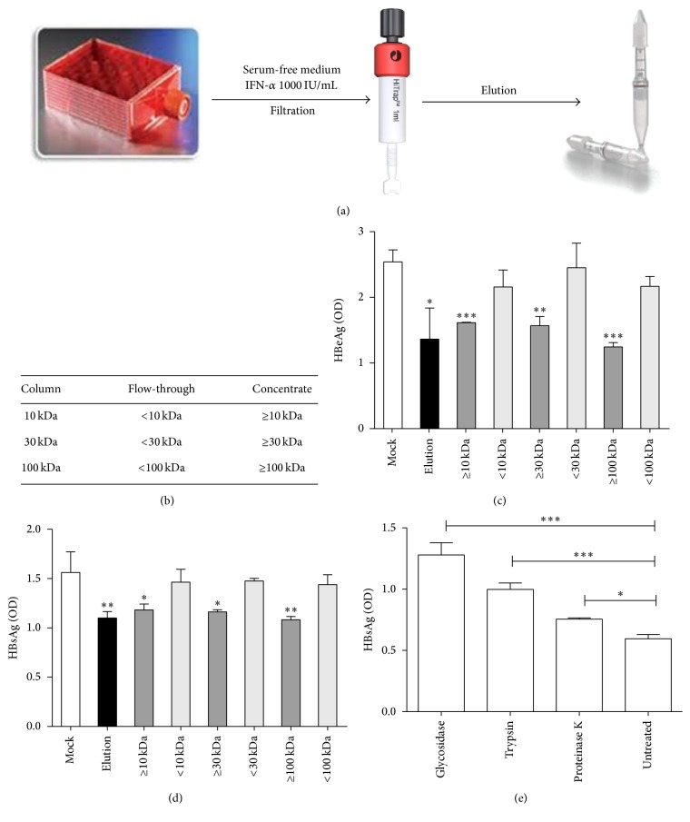
Figure 6.
Size exclusion of IFN-α induced binding inhibitors. (a) HepaRG cells were cultivated in hyperflask. After differentiation, cells were stimulated with 1000 IU/mL IFN-α for one day and then fed fresh serum-free medium. The heparin binding fraction was purified from heparin columns and protein concentration columns with different cutoffs were used to further separate the fractions by protein size. (b) Protein size of each fraction. (c) Differentiated HepaRG cells were incubated with indicated samples for 24 hours and then infected with HBV. HBeAg was evaluated by ELISA 4 days after infection. (d) Highly purified HBV SVPs from chronic HBV carriers were added to heparin-coated (25 μg/mL) 96-well plate and treated with different fractions. Plates were incubated for 2 h at 37°C, and heparin-bound SVPs were detected using an HBsAg ELISA kit. (e) Differentiated HepaRG cells were incubated with elution treated by glycosidase, or trypsin, or proteinase K for 24 hours and then infected with HBV. HBsAg was evaluated by ELISA 4 days after infection.

