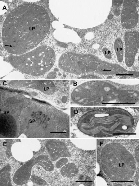
Figure 4.
Immunocytochemical localization of GPPS-SSU in peppermint. A, Immunogold labeling of GPPS-SSU (small black dots, arrows) in leucoplasts in a secretory cell of peltate glandular trichomes. B, Immunogold labeling of a freeze-substituted secretory cell leucoplast with anti-GPPS-SSU. C, Portion of a peltate gland stalk cell (below) and an adjacent secretory cell (above) labeled with anti-GPPS-SSU. Labeling is absent from stalk cell plastids. D, Representative chloroplast in mesophyll parenchyma of anti-GPPS-SSU treated section. The chloroplast is unlabeled. E, Preimmune treatment of secretory-stage secretory cells. Leucoplasts are unlabeled. F, A leucoplast of a preimmune treated secretory cell. LP, Leucoplast; SP, stalk cell plastid. Bars = 1 μm.

