Figure 2.
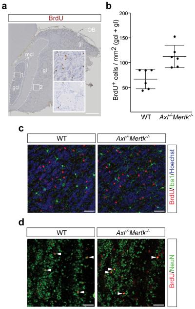
TAM signaling mediates ‘death by phagocytosis’. a, Axl−/−Mertk−/− OB section five weeks after BrdU pulse labeling, visualized with an anti-BrdU antibody (brown). The granule cell layer (gcl), glomerular layer (gl), and mitral cell layer (mcl) are indicated, and regions of the gcl (1) and gl (2) are enlarged. Scale bar 500 μm. b, Quantification of BrdU+ cells per mm2 in the gcl and gl of 6 WT versus 6 Axl−/−Mertk−/− mice; Graph plots average ± SEM; Two-tailed unpaired Mann Whitney p=0.002. c, BrdU+ cells in the gcl 35 days after injection of BrdU (red) are negative for Iba1 (green) in WT (left panel) and Axl−/−Mertk−/− (right panel) mice. d, Similar gcl sections stained with anti-BrdU (red) and NeuN (green). Arrowheads mark NeuN+BrdU+ cells. Panels in c co-stained with Hoechst 33258. Scale bars (c, d) 50 μm. Representative images of n = 2 per genotype.

