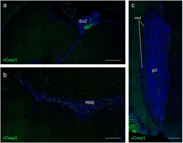
Extended Data Figure 2.
Apoptotic cell (AC) accumulation is confined to neurogenic and derivative migratory regions of the Axl−/−Mertk−/− CNS. a, A low power tiled image of a section through the Axl−/−Mertk−/− subventricular zone (SVZ) and surrounding brain tissue, stained for cCasp3, illustrates that ACs are confined within the SVZ. b, A low power tiled image of a section through the Axl−/−Mertk−/− rostral migratory stream (RMS) and surrounding brain tissue illustrates that cCasp3+ ACs are confined within the RMS. c, A low power tiled image of the granule cell and mitral cell layers (gcl and mcl, respectively) of the Axl−/−Mertk−/− olfactory bulb, stained for cCasp3, illustrates that there are no ACs detected in the double mutant bulb. Scale bars for a-c, 200 μm. Representative images from analyses performed in n = 3 mice (a-c)

