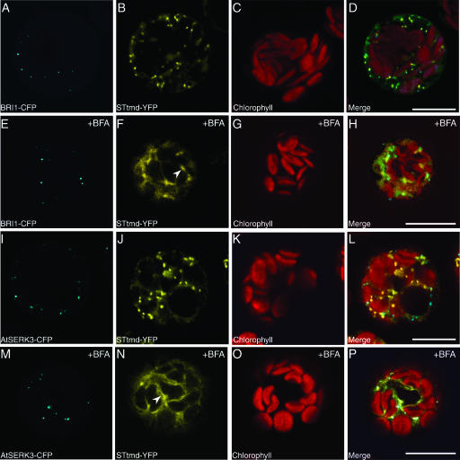
Figure 2.
Colocalization of BRI1 and AtSERK3 Proteins with the Golgi STtmd-YFP Marker in Cowpea Protoplasts.
Confocal images of protoplast cotransfected with BRI1-CFP/STtmd-YFP ([A] to [D]) and with AtSERK3-CFP/STtmd-YFP ([I] to [L]) recorded 3 h after transfection. The BRI1-CFP and AtSERK3-CFP fluorescence localized to the endosomes is shown in (A) and (I) (cyan). The STtmd-YFP fluorescence localized to the Golgi stacks is shown in (B) and (J) (yellow). The chlorophyll autofluorescence (red) is shown in (C) and (K) and the combined images in (D) and (L). Protoplast cotransfected with BRI1-CFP/STtmd-YFP ([E] to [H]) and with AtSERK3-CFP/STtmd-YFP ([M] to [P]) were allowed to express the proteins for 3 h and were then incubated in the presence of 20 μg/mL BFA for 30 min. The BRI1-CFP and AtSERK3-CFP fluorescence is shown in (E) and (M), respectively. The STtmd-YFP fluorescence is shown in (F) and (N). Membranes resembling the endoplasmic reticulum are indicated by arrowheads in (F) and (N). The chlorophyll autofluorescence is shown in (G) and (O) and the combined images in (H) and (P). Bars = 10 μM.

