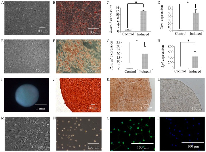
Figure 4.
Alizarin Red staining of SFMSCs prior to culture in osteogenic induction medium (A), and following culture in osteogenic induction medium for 4 weeks (B), and the expression of RUNX2 (C) and OCN (D) was compared between the osteogenic induction group and the control group. Oil Red O staining of SFMSCs prior to culture in adipogenic induction medium (E) and following culture in adipogenic induction medium for 4 weeks (F), and the expression of PPARG2 (G) and LPL (H) in the adipogenic induction group was compared to that in controls. Following culture in chondrogenic induction medium for 4 weeks, cartilage pellets formed in the tube (I), and the pellets were subjected to Safranin O staining (J) and immunohistochemical staining for type II collagen (K). The control group sections, which were treated using the same immunohistochemical staining process but without incubation with type II collagen antibody, exhibited negative staining (L). The morphology of SFMSCs prior to culture in neurogenic induction medium (M) and following culturing in neurogenic induction medium for 24 h (N); Cells after induction (O) were also stained for GFAP, while the control group (P) was not. Significant differences between the 2 groups are indicated by single asterisks (*p<0.05).

