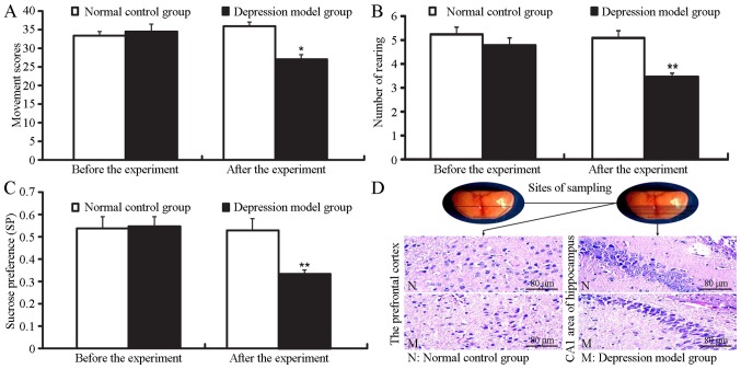
Figure 1.
Changes in behavior and histopathological changes in the prefrontal cortex and hippocampus in mice in the depression model group (means ± SEM). (A) Mobile scores; (B) number of rearing; (C) sucrose consumption. There was no distinctive difference between the two groups in the behaviors before the experiment (P>0.05). The mobile scores, number of rearing and sucrose intake of mice in the depression model group were significantly decreased compared with normal control group (P<0.05 or P<0.01); normal control group (n=21); depression model group (n=37); *P<0.05 or **P<0.01 compared with normal control group. (D) H&E staining (×200 magnification, n=4). In the normal control group, the cellular structures are compact with regular arrangement and a clear hierarchy in the prefrontal cortex and hippocampal CA1. In the depression model group, the morphology of the brain cells was changed from an ellipse or round shape to a spindle, strip or irregular shape. The cellular borders appeared fuzzy, and brain structures were disordered. Although the cellular arrangement of hippocampus CA1 region remained largely normal, the morphology of nerve cells changed significantly, and some nerve cells are absent in the depression model group.

