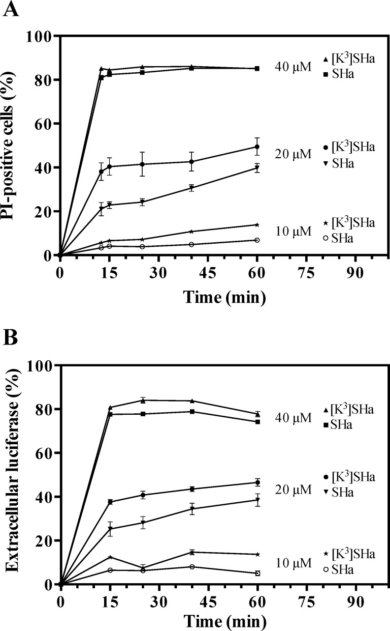
Fig 6.
Dose- and time-dependent propidium iodide (PI) staining (A) and luciferase release in the extracellular medium (B) of L. infantum parasites upon addition of temporins. L. infantum promastigotes (106 cells/ml) were incubated with different concentrations (10, 20 and 40 μM) of SHa or [K3]SHa for different times. PI-positive cells were counted by flow cytometry after adding PI (1 μg/ml) to the parasites. The luciferase activity in the extracellular medium was determined after centrifugation of the parasites and measurement of the luminescence using the Steady-Glo® Luciferase Assay System (Promega). The data are expressed as the means ± SEM of two experiments carried out in triplicate.

