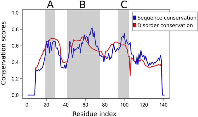
Fig 7. Sequence- and disorder conservation profile of CSDs.
The conservation of both the amino acid sequence (blue) and structural disorder (red) of CSD was quantified by considering a comprehensive set of homologous vertebrate CSD sequences covering mammals, birds and amphibians. The two anchoring regions (A and C) and the inhibitory segment (B) are shaded in grey.

