
Figure 3. Model of the trabeculation process.
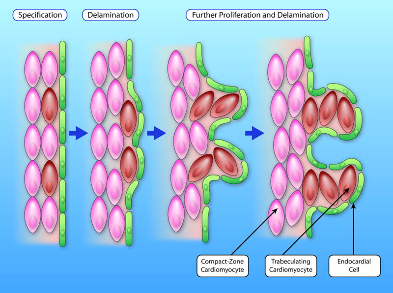
During trabeulation, a small fraction of CMs in the compact myocardium (pink) are first specified as trabeculating CMs (brown). These cells delaminate from the compact myocardium and migrate inward to form the first trabecular CMs. CMs in both compacted and trabecular myocardium further proliferate. This proliferation, together with CM migration and rearrangement, results in protrusion and expansion of the trabecular myocardium. (Illustration Credit: Ben Smith)