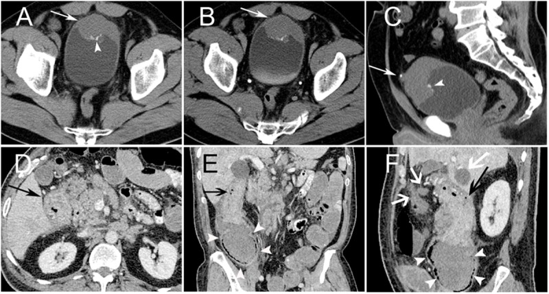
Fig 2. PNET Arising in the Intra-abdominal Region.

PNET of the bladder in a 54-year-old male (Case no. 1). Non-contrast CT image showed the anterior bladder wall with uneven thickening (arrow) and scattered punctuate calcification (arrowhead) (A). Contrast-enhanced CT showed mild homogenous enhancement of the mass (arrow) (B). Sagittal CT image showed calcification located on the mass surface (arrowhead), and a urachal stone was demonstrated (arrow) (C). Ascending colon PNET in a 65-year-old male (Case no. 12). CT images showed a locoregional ascending colon wall with uneven thickening and intermediate heterogeneous enhancement (black arrow) (D). Ascending colon locoregional luminal stenosis (arrow) (E, F) showed an incomplete intestinal obstruction with proximal colon dilatation (white arrowheads) (E). Retroperitoneal lymph node involvement and liver metastases were observed (white arrow) (F).
