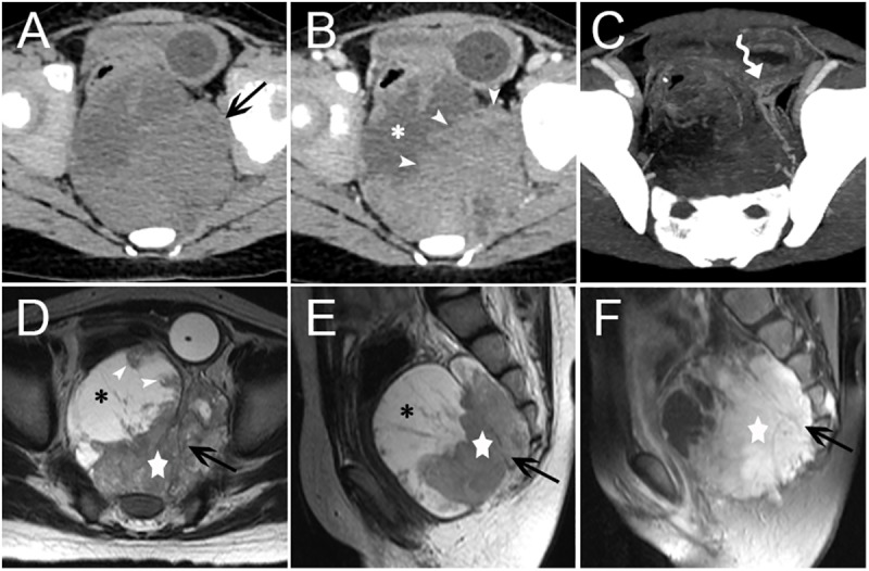
Fig 3. Presacral Region pPNET in a 5-year-old Female (Case no. 3).

CT images showed an irregular, mixed solid cystic mass (arrow) with an indistinct margin in the presacral region (A). The necrotic or cystic portion (asterisk) did not show clear enhancement, but the solid portion (arrowheads) presented with intermediate heterogeneous enhancement (B). Blood supply arteries could be observed (curved tail arrow) (C). Non-contrast MRI images showed the solid components (five-pointed star) of the mass (arrow) was isointense on T1WI images and mildly, heterogeneous hyper-intense on T2WI images. The lateral necrotic or cystic portion (asterisk) showed hypo-intensity on T1WI images and heterogeneous hyper-intensity on T2WI images. Mural nodules were observed (arrowhead) (D-F). On contrast-enhanced MRI images, the solid components (five-pointed star) of the mass showed marked heterogeneous enhancement, with low signal separation lines within the tumor (F).
