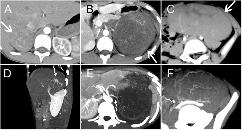
Fig 4. Tumor Blood Supply.

Three PNET cases with masses arising in the right adrenal gland region (A/D) (Case no. 2), left kidney (B/E) (Case no. 17) and mesentery (C/F) (Case no. 14) (arrows). Contrast-enhanced arterial phase CT images showed circuitous lines of small blood vessels within the masses. Tiny feeding arteries could be observed inside the mass, showing a crab-like appearance (curved tail arrow) (D-E).
